Single-neuron study finds signs of resilience in brain cells vulnerable to Parkinson's
Scientists sequenced mitochondrial DNA in individual brainstem neurons from people with Parkinson's for the first time, finding heavy damage but also a protective response that was stronger in people who lived longer.

Researchers at the University of Birmingham and Newcastle University have sequenced mitochondrial DNA in individual brainstem neurons from people with Parkinson's disease for the first time, the University of Birmingham said. The study, published on 7 October in the journal Brain, found extensive damage, along with signs that the cells fight back.
These neurons are linked to some of the hardest-to-treat symptoms
Most Parkinson's research focuses on dopamine-producing neurons, which are responsible for many of the movement symptoms the disease is best known for. This study looked at a different group: brainstem neurons that produce acetylcholine, a chemical messenger involved in sleep, thinking and movement.
Losing these neurons is associated with some of the most disabling and treatment-resistant symptoms, including problems with walking and balance, sleep disturbances, cognitive decline and, in some people, psychosis. The team studied neurons from post-mortem brain tissue donated by people with Parkinson's and compared them with healthy tissue.
Damaged cells ramp up a quality-control gene
The researchers found large-scale deletions in the neurons' mitochondrial DNA, especially in a region called the "major arc", which holds many genes needed to produce energy. Mitochondria act as the cell's power plants, so this kind of damage could leave neurons less able to cope with their workload.
But the affected neurons also had higher levels of PINK1, a gene that helps cells find and remove damaged mitochondria. Those levels were particularly high in neurons from people who had lived longer after their diagnosis, which may suggest a stronger quality-control response helps some neurons hold out for longer.
“Our findings reveal an extraordinary resilience within these vulnerable neurons. Although we observed extensive mitochondrial DNA damage, the cells appear to mount a protective response through increased PINK1 expression, likely as an attempt to maintain healthy mitochondria and preserve energy production.” — Dr Joanna Elson, Newcastle University, senior author
The work builds on earlier findings from the same researchers
Elson and co-senior author Dr Ilse Pienaar have studied these cells for years. In 2018, they reported mitochondrial DNA changes in cholinergic neurons of a brainstem region called the pedunculopontine nucleus, the University of Sussex said at the time. The new study is the first to analyse the DNA neuron by neuron.
The findings come from post-mortem tissue and show an association, not a treatment. The researchers hope they will lead to further studies of mitochondrial quality-control pathways as possible drug targets. Other recent work on nerve cells includes molecular threads that help neurons grow and form connections.
Like what you're reading?
Related stories
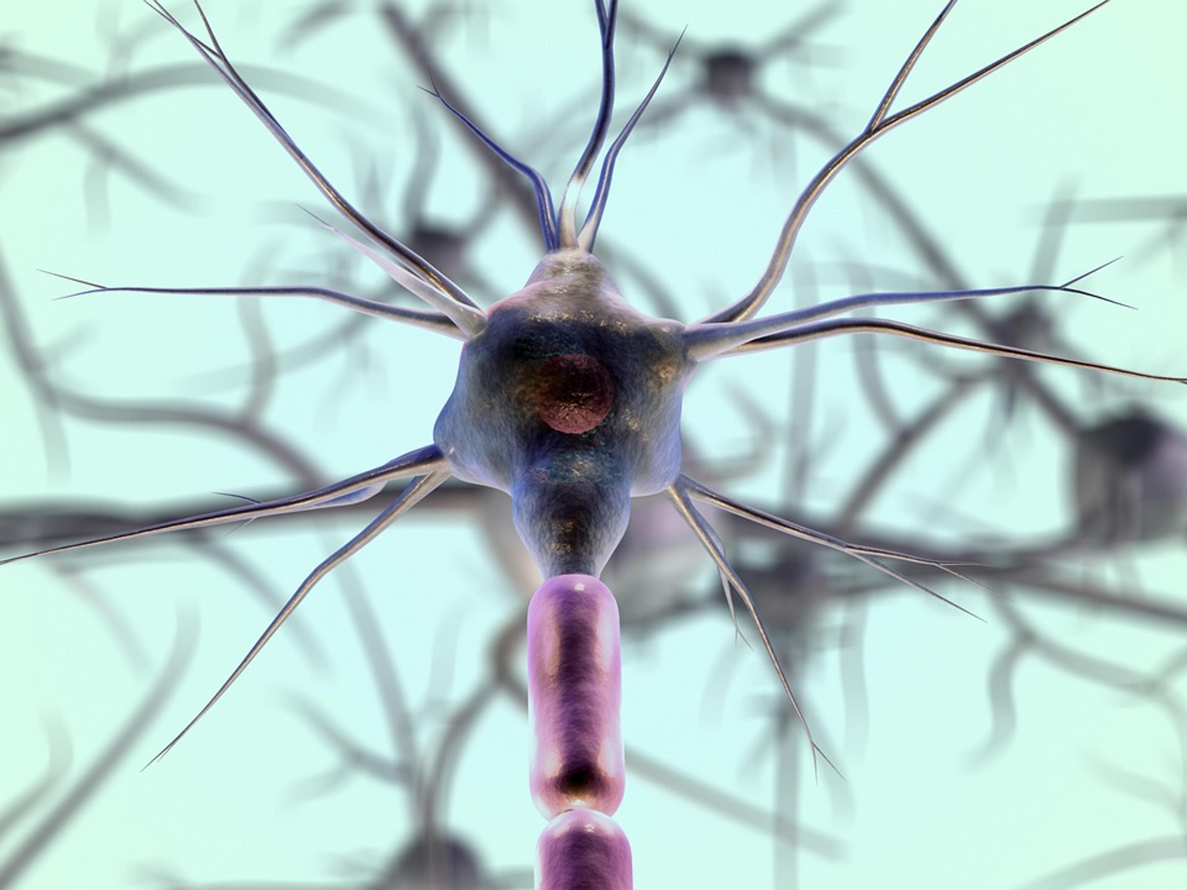
Scientists Built Molecular Threads That Quadrupled Neuron Activity in Lab Tests
Northwestern University researchers built self-assembling molecular threads that boosted neuron growth and synapse formation in lab tests, pointing toward future materials for brain and nerve repair.

NUS Researchers Partly Restore Egg Maturation in Stiff, Aged-Like Ovarian Tissue
National University of Singapore researchers found that stiffer ovarian tissue cuts the links between eggs and their support cells, and that blocking the protein SMAD7 partly restored egg maturation in lab-grown follicles from preclinical models.

Diabetes Drug Cut Inflammatory Blood Cells Within Two Weeks in Small Human Trial
In a Vanderbilt pilot study of 16 women with obesity and prediabetes, the SGLT2 inhibitor empagliflozin reduced inflammatory cell clumps linked to heart disease within two weeks, an effect a diet group did not show.